Vol 3 | Issue 2 | May-Aug 2017 | page: 13-14 | Yogen Bhatt, Girija Yadav, Smitha Rao.
Authors: M Sreedhar [1], Jaydeep Narendrabhai Anadkat [1], G V Chaitra [1], Kavyashree [1]
[1] Department of Anaesthesia, Adichunchanagiri Institute of Medical Sciences, BG Nagara, Karnataka, India.
Address of Correspondence
Dr. M Sreedhar,
Department of Anaesthesia,
Adichunchanagiri Institute of Medical Sciences,
BG Nagara – 571 448, Mandya, Karnataka, India.
E-mail: sreedharm83372@gmail.com
Abstract
Introduction: Plexiform neurofibromatosis is the hallmark lesion of neurofibromatosis Type 1. Neurofibromatosis is a type of benign tumor originating from the nerve sheath cells. Facial Neurofibroma can be extremely disfiguring.
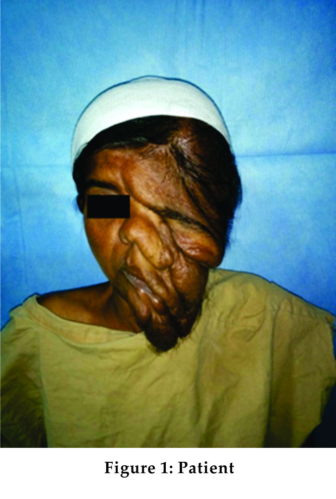
Case Presentation: A 43-year-old lady presented to our hospital for Epigastric hernia repair with distorted face because of a plexiform neurofibroma. The lesion involved the left half of the face.
Conclusion: The management of plexiform neurofibromatosis case, coming for other than corrective surgery, is challenging for the anesthesia team. Awake fiberoptic intubation can reduce the chances of perioperative complications related to difficult ventilation and intubation.
Keywords: Plexiform neurofibroma, epigastric hernia repair, difficult airway, ventilation.
Introduction
Neurofibromatosis is a type of benign tumor originating from the nerve sheath cells [1]. It is an autosomal dominant condition affecting about 1:2500-1:3000 live births [2]. Plexiform neurofibromas are the hallmark lesions of neurofibromatosis Type 1. They affect long portions of the nerve, sometimes infiltrating the nerve, and the surrounding tissue, thereby giving rise to an extensive disfiguration [3]. Here, we present a case of plexiform neurofibroma of the face coming for other than corrective surgery and the challenges we encountered while handling the case.
Case Report
A 43-year-old female presented to our hospital for epigastric hernia repair with the distorted face because of a plexiform neurofibroma. The lesion involved the left half of the face (Fig. 1). She had no other comorbidities. A history of delayed mile stones was noted. She weighed 35 kg, and her height was 154 cm. Her airway examination revealed Mallampati Class 3 and an inter-incisor gap of 3 cm. Neck extension was normal with adequate thyromental distance. Indirect laryngoscopy showed the presence of normal vocal cords. Spine examination revealed scoliosis. She was accepted for mesh repair under the American Society of Anesthesiologists (ASA) Grade 2 in view of anemia. In view of the anticipated difficult mask ventilation and intubation, thoracic epidural anesthesia was planned. An awake orotracheal fiberoptic intubation was prepared as alternative option. As part of the preparation, in the pre-operative area, she received 2% viscous lignocaine mouth gargle and nebulization with 4% lignocaine. Two 18-G peripheral cannulas were secured and she was briefed about the anesthesia plan. An attempt was made to place the epidural catheter in thoracic level, but was not successful due to presence of scoliosis. She was pre-medicated with 0.2 mg of glycopyrrolate, intravenously. In the operation theater, awake intubation was facilitated by performing bilateral superior laryngeal nerves and transtracheal recurrent laryngeal nerve block with 2 ml of 2% lignocaine each combined with spraying the vocal cords with 2% lignocaine. Fiberscope was negotiated through the vocal cords. The awake intubation process was uneventful. The position of endotracheal tube was confirmed by capnogram and auscultation with stethoscope. The patient was induced with thiopentone (250 mg) and vecuronium (3.5 mg). Anesthesia was maintained with oxygen (O2), nitrous (N2O), and isoflurane. Fentanyl and vecuronium were supplemented intermittently. After surgical procedure reversal agent (neostigmine 1.75 mg + glycopyrrolate 0.35 mg) was given. Extubation was uneventful. She was shifted to the recovery room for observation. She was receiving supplemental O2 through Hudson’s mask at 4 L/min for 2 h. There was minimal soakage of the dressing. Her vital parameters were normal. She was discharged after 5 days of post-operative stay.
Discussion
A neurofibroma is a nerve sheath tumor occurring in the peripheral nervous system, inherited as an autosomal dominant trait. They can result in a range of symptoms from physical disfiguration and pain to cognitive disability. Neurofibromas have been subdivided into two broad categories: dermal and plexiform. Dermal neurofibromas are associated with a single peripheral nerve while plexiform neurofibromas are associated with multiple nerve bundles [4]. Plexiform neurofibroma is generally considered a hallmark of Type 1 neurofibromatosis. Airway assessment is important when it involves the face and the oropharyngeal structures. Intraoral manifestation is seen in around 5% of patients [5]. Plexiform neurofibromas can undergo malignant transformation. In addition to the careful airway assessment, a thorough systemic examination is equally important. Pheochromocytoma and renal artery stenosis can be associated with plexiform neurofibromatosis. In the presence of pre-operative hypertension, these causes should be looked into. It is important to discuss the anesthesia plan, the need for blood transfusion, post-operative intensive care unit care, and prolonged hospitalization. An awake intubation was done in our case anticipating difficult mask ventilation and intubation. A difficult airway cart should be easily accessible. Patients with neurofibromatosis may have associated coagulation abnormality [6]. This case highlights the limitations of ASA grading. It offers an inadequate distinction between the presence of a systemic illness and localized manifestation of a systemic illness. It also fails to incorporate the airway grading. A difficult airway may predispose to increased risks for the patient. In addition, the grading system does not include the nature of surgery, which by itself, is a predictor of morbidity and mortality.
Conclusion
The management of plexiform neurofibromatosis case, coming for other than corrective surgery, is challenging for the anesthesia team. The major challenges for this case were plan of anesthesia and preparation for it. Awake fiberoptic intubation can reduce the chances of perioperative complications related to difficult ventilation and intubation.
References
1. 1. Kim MJ, Cheon CK. Neurofibromatosis Type 1: A single center’s experience in Korea. Korean J Pediatr 2014;57:410-5.
2. Darrigo LG Jr, Geller M, Filho AB, Azulay DR. Prevalence of plexiform neurofibroma in children and adolescents with Type 1 neurofibromatosis. J Pediatr (Rio J) 2007;83:571-3.
3. Hirsch NP, Murphy A, Radcliffe JJ. Neurofibromatosis: clinical presentations and anaesthetic implications. Br J
Anaesth 2001;86:555-64.
4. Lan K, Christian H, Victor M. Nervous system: Neurofibroma. Atlas Genet Cytogenet Oncol Haematol 2007;11:344-7.
5. Ali QE, Amir SH, Shafi M, Chaudhri TR. Awake airtraq intubation in plexiform neurofibroma of face: A new
experience. Indian J Anaesth 2013;57:97-8.
6. Vélez R, Barrera-Ochoa S, Barastegui D, Pérez-Lafuente M, Romagosa C, Pérez M, et al. Multidisciplinary management of a giant plexiform neurofibroma by double sequential preoperative embolization and surgical resection. Case Rep Neurol Med 2013;2013:987623.
| How to Cite this Article: Sreedhar M, Anadkat JN, Chaitra GV, Kavyashree. Anesthetic challenges of plexiform neurofibroma of the face with epigastric hernia for repair. Journal of Anaesthesia and Critical Care Case Reports May-Aug 2017;3(3):13-14. |